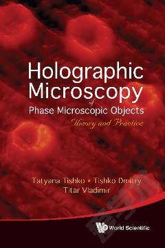
Holographic Microscopy Of Phase Microscopic Objects: Theory And Practice
The book presents a clear and comprehensive review of the current status of the holographic microscopy with discussion of the positive and negative features of classical and holographic methods for solving the problem of three-dimesional (3D) imaging of phase microscopic objects. Classical and holographic methods of phase, interference and polarization contrast are discussed. Combination of the developed holographic methods with the methods of digital image processing allowed creating the digital holographic interference microscope (DHIM). The first 3D images of native phase microscopic objects such as blood cells were obtained using the DHIM. The results of DHIM application for study of blood erythrocytes, thin films, micro-crystals are presented.
{{comment.content}}


 京公网安备 11010802027623号